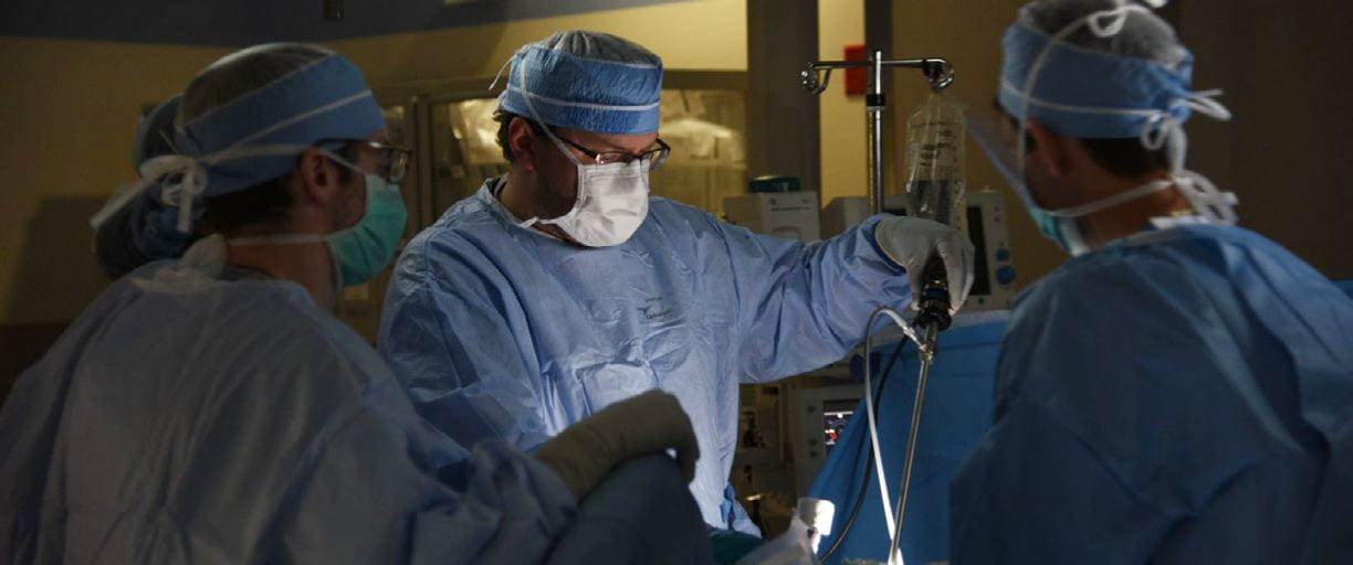
Washington University Colorectal Surgeons answer patient questions regarding rectal cancer. As internationally recognized leaders in the field, Washington University surgeons partner with Siteman Cancer Center to treat about 350 new colorectal cancer patients a year.

The rectum is the last 6-8 inches of the colon. The cells that develop into cancer are the same in the colon and the rectum. However, there are differences between the colon and the rectum that impact how their respective cancers are treated.
The differences include function and location. The main function of the colon is to absorb water and transport stool to the rectum. The rectum’s main function is to store stool until deemed appropriate for a bowel movement. Anatomically, the rectum coursed from the peritoneal cavity through the rectum to the anus. It is because of the its location in the pelvis and the fact that it is located outside of the peritoneal cavity that rectal cancer is treated differently than colon cancer
Rectal cancer is staged clinically to help determine the specific treatment that the patient will receive. The first step is for your surgeon to examine your rectum to determine the exact location of the tumor. This is done with a digital rectal examination and proctoscopy. Proctoscopy is performed by inserting a scope into the rectum to determine if the tumor is in the upper or lower rectum. This is easily done in the office and does not require sedation.
A CT scan of the chest and abdomen is used to detect if there is tumor that has spread outside of the rectum. A specialized MRI of the pelvis is used to stage the primary tumor in the rectum. The MRI is able the determine how far through the wall of the rectum the tumor has grown, how close it is to surrounding structures and if there are any lymph nodes involved. Together these studies give the clinical stage of the tumor.
The pathologic or final stage is obtained once the rectum has been removed and the pathologist examines the tumor and lymph nodes under a microscope.
Rectal cancer is treated based on its clinical stage or the results of the pelvic MRI and CTs of the chest and abdomen. Typically, rectal cancers that are contained within the wall of the rectum and do not have any detectable lymph nodes are treated with surgery alone and the need for chemotherapy is determined based on the pathologic stage.
Rectal cancers that have grown through the wall of the rectum and/or have detectable lymph nodes are treated with chemotherapy and radiation treatments before surgery. Historically, the radiation therapy consisted of 25-28 treatments over a 5-6 week period. The chemotherapy was used to increase the effectiveness of the radiation therapy and not necessarily kill tumor cells. Surgery then occurred 8-10 weeks later after all of the inflammation of the radiation resolved. Then, based on a combination of the clinical and pathologic stage, the need for systemic chemotherapy was determined. This treatment regimen was long and intense, lasting up to 44 weeks. It was very effective at preventing the tumor from returning in the pelvis but did not decrease the risk of the tumor showing up somewhere outside of the rectum.
As are result, we at Washington University have made two major adjustments. First, we use what is called short course radiation therapy. The same biologic dose of radiation is delivered in 5 days rather than 5 weeks. Second, we moved the timing of the systemic chemotherapy from after surgery to before surgery. Thus, the order of treatment is 5 days of radiation therapy, 2-week rest period, 24 weeks of chemotherapy, 4-week rest period and then surgery. This regimen is called total neoadjuvant therapy (TNT), as all chemotherapy and radiation are given before surgery. The total treatment time with this regimen is typically 31 weeks compared to 44 weeks with the other regimen.
There are several benefits to TNT:
1) Shorter overall treatment time
2) The systemic chemotherapy is better tolerated before surgery than after surgery
3) Patients receive more systemic chemotherapy when given before than after surgery
4) More total patients receive systemic chemotherapy
5) The rectal cancer is more likely to shrink
6) Early results indicate that these patients have a better chance of survival
The surgery for rectal cancer entails removal of the sigmoid colon, rectum and associated lymph nodes. The technical aspects of this are very important. It is necessary to remove the colon, rectum and lymph nodes all within the embryonic package in which they are contained. The key components are to obtain adequate margins above, below and around the tumor. The extent of the rectum removed is dependent upon where in the rectum the tumor is located. Typically, tumors that do not involve the sphincter muscles can be removed without the need for a permanent colostomy.
For tumors above the anal sphincter muscles, the tumor is removed though the abdomen, and the colon is connected to the remaining rectum. This is called a low anterior resection. A major risk of the operation is if that connection between the colon and rectum were to leak or not heal completely, which occurs about 5% of the time. To minimize the risk of this happening and to be able to salvage the connection if it were to happen, often a temporary stoma or bag is created. The colon and rectum are connected, but stool leaves the intestine through the stoma. Nothing passes by the connection, thus protecting it. The stoma typically remains in place for 3 months or until any remaining chemotherapy is completed. Because the colon and rectum have already been connected, removing the temporary stoma is a much easier operation than the original surgery.
Tumors that are growing into anal sphincter muscle require removal of the pelvic floor and permanent colostomy. This operation is called an abdominoperineal resection (APR). The technical steps are the same, except to remove the pelvic floor and anus an incision is made. In addition to going through the abdomen, there is an incision in the perineum or bottom where the anus and sphincter have been removed.
Surgery remains a mainstay in the treatment for rectal cancer.
However, as our understanding of the effects of chemotherapy and radiation on the rectal cancer improves, we are learning that selected patients may be able to avoid an operation. Patients who receive chemotherapy and radiation as the first steps of their treatment may be considered for management without an operation. This is dependent upon the extent of the response of the tumor to the chemotherapy and radiation. Roughly, 1 in 4 patients will have what we call a complete pathologic response. This is the group of patients that can be managed without an operation.
The key is being able to identify those patients. After completion of the radiation and chemotherapy, patients are restaged with an MRI, digital rectal exam and sigmoidoscopy. These three studies are used together to determine the response to the treatment and identify the presence or absence of residual tumor. Patients with residual tumor are recommended to undergo an operation. Patients with no clinical evidence of remaining tumor can then be watched closely. The watching or surveillance consists of imaging with either a CT scan or MRI, digital rectal exam and sigmoidoscopy every 3 months for the first 2 years. If regrowth of the tumor is detected during surveillance, then an operation is needed. Typically, if the tumor is going to return it occurs within the first 2 years.
Low anterior resection syndrome is a collection of symptoms or issues patients have after undergoing a resection or removal of part or the entire rectum. These symptoms may include:
· frequency/urgency of stools
· clustering of stools (numerous bowel movements over a few hours)
· stool incontinence
· no stool for a day or two or more and then numerous bowel movements another day
· increased gas
Not all patients experience every symptom and each patient is unique.
Typically, symptoms are worse right after surgery and improve up to 2 years after surgery. The reasons for the symptoms are multifactorial and related to the function of the rectum and colon. The colon functions to absorb water from the stool and move it to the rectum, where it is stored until the patient is ready for a bowel movement. When a patient has undergone a low anterior resection, the majority of the rectum has been removed, so there is less room to store stool. As a result, it takes less stool to accumulate in the rectum to signal the brain that it needs to be emptied. This is what leads to the frequency and clustering of bowel movements.
The other issue is the urgency and potential seepage of stool. Normally the internal and external sphincter work together to maintain fecal continence. The internal sphincter is a muscle which we do not control, but we have complete control of the external sphincter. Before surgery, when there is an urge to have a bowel movement, the sensation begins and it is then determined if this is an appropriate time and place for a bowel movement. If it is not appropriate to have a bowel movement, we hold it and eventually that sensation goes away until we find the appropriate time. The internal sphincter muscle is responsible for this reflex. After surgery, when the rectum has been divided, this reflex goes away. This means that control of the bowels is mostly dependent upon the external sphincter – the muscle we control. Like any other muscle that we control, it fatigues, so that initial sense of needing to have a bowel movement progresses to urgency as the muscles fatigue.
The goal of treatment for LARS is improved bowel function so that patients are able to have the best possible quality of life. There are several aspects that are involved in improving a patient’s bowel function. First, education is extremely important.
1. Muscle strengthening exercises combined with dietary changes may help with urgency and stool incontinence.
2. For clustering of bowel movements try:
1. Imodium AD: You may start by taking one 2 mg tablet prior to each meal, increasing to two tablets 4 times a day. You may take up to 16 mg of Imodium daily. Imodium should be taken before loose bowel movements, ideally 30 minutes before meals and at bedtime.
2. A probiotic such as FloraQ, Align or VSL #3 (available online) may be helpful.
3. Citrucel or Metamucil, one dose in a glass of water or juice at bedtime
3. Chew foods thoroughly.
4. Try small, frequent meals (5-6 per day). Skipping meals may worsen watery stools and cause increased gas.
5. Add new foods one at a time to determine the effect each has on your bowel movements.
6. Drink plenty of fluids. Sip fluids slowly and drink either between meals or at the end of a meal.
7. Avoid caffeine and/or alcohol. These can worsen stool output.
8. Eat foods high in soluble fiber and use fiber supplements. Psyllium-based products improve stool consistency by absorbing water but not reducing the volume. This may help slow and thicken the stool.
9. Milk and milk products contain lactose and can worsen diarrhea for some people. Try lactose free milk or enzyme tablets if milk affects you.
10. Imodium AD is an anti-diarrheal medication that is available over the counter that is well-tolerated and may improve anal sphincter pressure. This helps thicken stool as well as helps with stool incontinence.
11. Carry a “survival pack” consisting of wet wipes, protective ointments (example: Calmoseptine or other “diaper-type” barrier ointments), and Imodium AD.
The goal of treatment for LARS is improve a patient’s bowel function so that they are able to have the best quality of life as possible. The only indication for surgery is when a patient’s quality of life
is such that they would rather have permanent colostomy. Therefore, the only surgery available for LARS is the removal of the remaining rectum and the creation of a permanent colostomy.
Washington University Colorectal surgeons outlines which foods can cause gas.
Foods that can cause gas:
- Cabbage
- Dairy products
- Brussels sprouts
- Spinach
- Broccoli
- Radishes
- Cauliflower
- Carbonated beverages
- Onions
- Beans
- Corn
- Cucumbers
- Nuts
- Beer
Washington University Colorectal surgeons outlines which foods can make stool firmer and more regular.
Foods that make stools firmer:
- Bananas
- White boiled rice
- White pasta
- White bread (not high fiber)
- Milk
- Arrowroot biscuits
- Marshmallows (white)
- Tapioca
- Peanut butter
- Potatoes
- Cheese
- Yogurt
- Pretzels
Foods that may cause softer and more frequent stools:
- Vegetables: red capsicum, cabbage, onions, spinach, dried and fresh beans, peas, corn, Brussels sprouts and broccoli
- Fruit: Fresh, canned or dried fruit; grapes, apricots, peaches, plums and prunes
- Spices: Chili, curry and garlic
- Caffeine: Coffee, tea, cola and chocolate
- Alcohol: beer and red wine
- Glucose-free foods containing Sorbitol or Mannitol: Sugar-free chewing gum, some mints, sweeteners and snack bars
- Bran, other high fiber cereals and breads as well as some fiber supplements
- Milk and other dairy products
- Nuts and popcorn
- Greasy foods
- Prune, orange and grape juice
Washington University Colon and Rectal can provide screening, care options and treatments for colorectal cancer. Meet our specialists below.


